Researchers hope that studying these organoids would aid them in the development and testing of new treatments for pancreatic cancer, which is one of the deadliest types of disease.
Engineers at the Massachusetts Institute of Technology (MIT), in partnership with scientists at the Cancer Research UK Manchester Institute, have created a new method of growing small replicas of the pancreas, which can be made from either healthy or malignant cells. Scientists hope that their new models may aid them in the development and testing of possible treatments for pancreatic cancer, which is currently one of the most difficult types of cancer to cure.
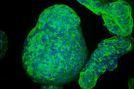
The researchers were able to develop pancreatic “organoids” in a customised gel that mimicked the extracellular environment surrounding the pancreas,
allowing them to explore the crucial interactions between pancreatic cancers and their surrounding environment. To distinguish it from some of the gels already in use for tissue growth, the new MIT gel is totally synthetic, simple to build, and can be made with a consistent composition every time.
According to Linda Griffith, the School of Engineering Professor of Teaching Innovation and a professor in both biological engineering and mechanical engineering, “reproducibility is a significant concern.” In order to undertake more methodical cultures of these kinds of organoids, and in particular to regulate the microenvironment, the research community has been exploring for ways to do so.”
Researchers have also demonstrated that their novel gel may be used to produce different forms of tissue, including intestinal and endometrial tissue, according to their findings.
Among the senior authors of the work, which appears today in Nature Materials, are Griffith and Claus Jorgensen, a group leader at the Cancer Research UK Manchester Institute in Manchester, England. Among the authors is Christopher Below, a former graduate student
at the Cancer Research UK Manchester Institute who is now working as a freelance writer.
Creating a microenvironment that is similar to the real one
Organoids grown in a lab dish have traditionally been grown using commercially available tissue-derived gel, which may be purchased online. In spite of this, Griffith notes that because the most extensively used commercial gel is a complicated mixture of proteins, proteoglycans, and growth factors produced from a tumour established in mice, it is varied from lot to lot and may contain unwanted components. It also does not always allow for the proliferation of a variety of cell types to coexist. It was about ten years ago that Griffith’s lab began working on developing a synthetic gel that could be used to grow epithelial cells, which form the sheets that line the inside of most organs, as well as other supportive cells in culture.
The gel that they developed is based on polyethylene glycol (PEG), a polymer that is frequently utilised in medical applications since it does not interact with living cells in the same way that other materials do. In order to better understand the biochemical and biophysical properties of the extracellular matrix, which is the layer of tissue that surrounds organs in the body, the researchers studied the extracellular matrix and identified characteristics that could be incorporated into the PEG gel to aid in the growth of cells in it.
One distinguishing characteristic is the presence of small molecules known as peptide ligands, which interact with cell surface proteins known as integrins. Cells cling to the gel and form organoids as a result of the sticky binding that occurs between ligands and integrins. Small synthetic peptides generated from fibronectin and collagen were added to the gels, and the researchers discovered that this allowed them to produce a range of epithelial tissues, including intestinal tissue. Additionally, they demonstrated that supporting cells known as stromal cells, in addition to immune cells, can flourish in this environment.
Griffith and Jorgensen wanted to investigate if the gel could be utilised to support the growth of normal pancreatic organoids as well as pancreatic cancers in the latest investigation. As a result, it has traditionally been difficult to develop pancreatic tissue in a way that mimics both the malignant cells and the surrounding environment, as pancreatic tumour cells lose their dangerous characteristics once they have been removed from the body.
A process for producing the new gel was created by Griffith’s group, which then collaborated with Jorgensen’s lab, which investigates the biology of pancreatic cancer, to put it through its paces. Jorgensen and his students were able to create the gel and utilise it to develop pancreatic organoids from either healthy or malignant pancreatic cells taken from mice, according to the researchers.
It was a simple process once we received the protocol from Linda and placed the reagents in the system, according to Jorgensen. “I believe that says volumes about how robust the system is, as well as how simple it is to put it into practise in the lab.”
He claims that other procedures they had explored were either too complicated or did not accurately replicate the microenvironment found in real tissues. When Jorgensen’s lab used this gel to compare the pancreatic organoids to tissues that they had previously analysed in real mice, the researchers discovered that the tumour organoids expressed a number of the same integrins as were identified in pancreatic tumours. Other types of cells that often surround tumours, such as macrophages (which are immune cells) and fibroblasts (which are supporting cells), were also able to thrive in the tumour microenvironment.
Cells obtained from patients
Aside from this, the researchers demonstrated that they could utilise their gel to create organoids from pancreatic cancer cells obtained from patients. They believe it could also be valuable in the research of cancers such as lung, colorectal, and other types. Such systems could be used to investigate the effects of putative cancer medicines on tumours and their surrounding microenvironment.
Furthermore, Griffith intends to utilise the gel to grow and study tissue obtained from patients suffering from endometriosis, a disorder in which the tissue that lines the uterus begins to proliferate outside of the body. This might cause discomfort and, in some cases, infertility.
Its synthetic nature means that it can be easily created in a laboratory by combining specified precursors, such as PEG and certain polypeptides. This is one of the advantages of the new gel. According to the researchers, they have filed a patent application for their invention and are currently in the process of licencing it to a firm that could manufacture the gel on a commercial scale.
In addition to Cancer Research UK and the Rosetrees Trust, the research was supported by a European Research Council Consolidator Award as well as funding from the National Science Foundation. The National Institutes of Health and the Defense Advanced Research Projects Agency provided additional support.
For More Information Visit : Theny Magazine